

Taser Cardiac Monitoring of Subjects Exposed to Taser Ucsd Emergency Medicine
Download original document:
Document text
Document text
This text is machine-read, and may contain errors. Check the original document to verify accuracy.
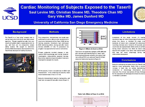
Cardiac Monitoring of Subjects Exposed to the Taser®
Saul Levine MD, Christian Sloane MD, Theodore Chan MD
Gary Vilke MD, James Dunford MD
University of California San Diego Emergency Medicine
Background

500

Methods

Limitations

400
300

The Taser® is a “less lethal” weapon now in
use by nearly one third of U.S. law enforcement
agencies. Though regarded as safe, there are
reports of sudden death in association with its
use, and there are no prospective human
safety studies. We sought to determine
whether ECG disturbances developed when the
Taser® was deployed on healthy volunteers.

This prospective, interventional pilot study was
performed on police officers who volunteered to
experience deployment of the Taser X26® during
training sessions. Volunteers had continuous
ECG monitoring before, during and after shock
delivery (50,000V, 2.1 mA, 19 pulses/sec) via
alligator clips applied to the trunk or extremities.
Primary endpoints included changes in rate,
rhythm, ECG morphology and interval duration.
Investigators independently analyzed tracings.
Descriptive statistics & paired student t test were
employed to evaluate the results.

ms

0
PR

QRS

Figure 2: Effect of shock on ECG

post-

pre-

post-

pre-

118

138

149

146

69

67

431

441

112125

134144

142157

136155

6374

6274

415447

424458

100Range 66-160 94-190 210

100240

40120

50120

350620

283580

∆ HR
(bpm)

∆ PR
(ms)

∆ QRS
(ms)

∆ QTc
(ms)

<0.0001

0.22

0.35

0.19

-24.6 to
-14.5

-2.25 to
9.47

-1.7 to
4.7

-26.4 to
5.5

-60 to
40

-30 to
20

-110 to
145

135
130

Range -1 to 72

138

125
118

Table 1a/b: Effect of Taser ® on ECG

110
105

POST-SHOCK

Limitations of this study include: (1) healthy
volunteers may not reflect physiology of subjects for
whom this device is intended (2) single brief
durations of shock were employed (3) post-shock
recording intervals were limited to 30 seconds and
could miss late asymptomatic ECG events (4) artifact
during shock obscures true ECG (5) shock was
applied with alligator clips rather than barbed darts
and may not mimic physiology during law
enforcement deployment.

Conclusions

post-

95%
C.I.

Pvalue
95%
C.I.

PRE-SHOCK

pre-

Mean

Subjects demonstrated baseline tachycardia, and
heart rate increased 20 bpm after shock (Figure 1).

HR
120
(bpm)
115

post-

HR
HR PR PR QRS QRS QTc QTc
(bpm) (bpm) (ms) (ms) (ms) (ms) (ms) (ms)

Of 58 subjects, 9 were excluded due to ECG lead
displacement. In the remaining 49 subjects, mean
shock duration was 2.3 sec (1.2– 5.0).

140

QTc

There were no significant changes in PR, QRS and
QTc intervals post Taser® (Figure 2, Table 1a/b).
Sinus tachycardia was common; one subject had
rare unifocal PVC’s pre and post Taser®. Artifact
obscured rhythm during shock (Figure 3)

Results

Researcher physician enjoying Taser ®
with police training staff

POST

100

pre-

Taser X-26®

PRE

200

Subjects undergoing voluntary shock by Taser® had
baseline sinus tachycardia. Aside from a further
increase in heart rate, no cardiac dysrhythmia,
conduction or morphology disturbances were
observed. The clinical implications of these findings
for the population of individuals for whom this device
is intended is unknown.

Disclosures

This study was accomplished without outside funding

Acknowledgements
Special thanks goes to the San
Diego Police Department for
allowing this study to be performed
during scheduled officer training and
to the San Diego Fire Rescue
Department for providing ECG