
Taser Physiological Effects of Elec Weapon on Human Subj Annals Emerg Medicine 2007
Download original document:
Document text
Document text
This text is machine-read, and may contain errors. Check the original document to verify accuracy.
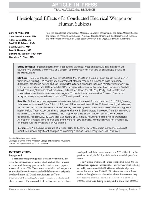
ARTICLE IN PRESS TRAUMA/ORIGINAL RESEARCH Physiological Effects of a Conducted Electrical Weapon on Human Subjects Gary M. Vilke, MD Christian M. Sloane, MD Katie D. Bouton, BS Fred W. Kolkhorst, PhD Saul D. Levine, MD Tom S. Neuman, MD Edward M. Castillo, PhD, MPH Theodore C. Chan, MD From the Department of Emergency Medicine, University of California, San Diego Medical Center, San Diego, CA (Vilke, Sloane, Levine, Neuman, Castillo, Chan); and the Department of Exercise and Nutritional Sciences, San Diego State University, San Diego, CA (Bouton, Kolkhorst). Study objective: Sudden death after a conducted electrical weapon exposure has not been well studied. We examine the effects of a single Taser exposure on markers of physiologic stress in healthy humans. Methods: This is a prospective trial investigating the effects of a single Taser exposure. As part of their police training, 32 healthy law enforcement officers received a 5-second Taser electrical discharge. Measures before and for 60 minutes after an exposure included minute ventilation; tidal volume; respiratory rate (RR); end-tidal PCO2; oxygen saturation, pulse rate; blood pressure (systolic blood pressure/diastolic blood pressure); arterialized blood for pH, PO2, PCO2, and lactate; and venous blood for bicarbonate and electrolytes. Troponin I was measured at 6 hours. Data were analyzed using a repeated-measures ANOVA and paired t tests. Results: At 1 minute postexposure, minute ventilation increased from a mean of 16 to 29 L/minute, tidal volume increased from 0.9 to 1.4 L, and RR increased from 19 to 23 breaths/min, all returning to baseline at 10 min. Pulse rate of 102 beats/min and systolic blood pressure of 139 mm Hg were higher before Taser exposure than at anytime afterward. Blood lactate increased from 1.4 mmol/L at baseline to 2.8 mmol/L at 1 minute, returning to baseline at 30 minutes. pH And bicarbonate decreased, respectively, by 0.03 and 1.2 mEq/L at 1 minute, returning to baseline at 30 minutes. All troponin I values were normal and there were no EKG changes. Ventilation was not interrupted, and there was no hypoxemia or hypercarbia. Conclusion: A 5-second exposure of a Taser X-26 to healthy law enforcement personnel does not result in clinically significant changes of physiologic stress. [Ann Emerg Med. 2007;xx:xxx.] 0196-0644/$-see front matter Copyright © 2007 by the American College of Emergency Physicians. doi:10.1016/j.annemergmed.2007.05.004 INTRODUCTION Background There has been growing public demand for effective, less lethal law enforcement weapons, which include blunt impact weapons such beanbag guns or rubber bullets, mace, pepper spray, and batons. The Taser, a conducted electrical weapon, is an electrical law enforcement and self-defense device originally developed in the 1970s and manufactured by Taser International (Scottsdale, AZ). Early versions were bulky and often ineffective. Various models of the Taser device have been Volume xx, . x : Month developed, and their newest version, the X26, differs from the previous model, the M26, mainly in the size and shape of the device. The National Institute of Justice reports that 9,800 US law enforcement agencies authorize the Taser device, which is being carried by more than 225,000 officers.1 Additionally, they report that more than 120,000 US citizens also have a Taser device. Although the actual number of uses is unknown, they have reported that the Taser has been used on more than 150,000 volunteers during training and in more than 100,000 Annals of Emergency Medicine 1 ARTICLE IN PRESS Physiological Effects of Conducted Electrical Weapons on Humans Editor’s Capsule Summary What is already known on this topic The Taser delivers an electrical pulse, causing incapacitating tetanic muscular contractions. Sudden death has been associated with its use. What question this study addressed What are some measurable physiologic effects of a single 5-second Taser exposure on healthy human volunteers? What this study adds to our knowledge In 32 healthy individuals, there were no clinically important changes in respiratory, cardiac, acid-base, or electrolyte status after a single 5-second Taser exposure. How this study might change clinical practice Although this small study suggests that the Taser is safe in healthy volunteers, there are insufficient data to determine its safety profile in individuals with agitated delirium, the population on whom the device is mainly used. “real-life” police confrontations. The manufacturer asserts that the device helps officers avoid the use of deadly force while lowering the risk of injury to officers. The Taser X26 is designed to be deployed up to 7 m from the subject. The operator fires the device, releasing 2 9-mm barbs attached to the gun by thin, 7-m copper wires. When the circuit is completed, an electrical pulse of 5 seconds’ duration is automatically delivered through the wires to incapacitate the subject by causing involuntary tetanic muscular contractions. The officer may deliver continued electricity by pulling the device trigger again. Although the effect of the Taser is poorly studied, it is generally regarded as safe2-4 and has been approved by the US Consumer Product Safety Commission for the current indication for which it is being used. Most of the data supporting the product’s approval by the US Consumer Product and Safety Commission was based on theoretic calculations and not animal or human studies.5 Importance There have been a number of reports of sudden death after Taser administration. Amnesty International6 reports “152Taser related deaths” since 2001, and the Arizona Republic7 reports “167 cases of death following stun gun use” since 1999. The majority of deaths in humans who were exposed to a Taser device were associated with illicit drug use, especially phencyclidine, methamphetamine, and cocaine.4,8,9 However, there have been several deaths reported in individuals after Taser exposure who were not under the influence of illicit drugs. These cases generally involved a clinical presentation of “excited delirium” and other comorbid factors that were likely to be 2 Annals of Emergency Medicine Vilke et al related as the cause of the suspect’s death.10-12 Most case reports and police reports note that such suspects who are shocked with a Taser go into cardiac arrest 5 to 40 minutes later.13 If a lethal dysrhythmia, particularly ventricular fibrillation, was at fault from the electrical discharge, cardiac arrest would be expected to occur at the Taser activation. However, if individuals were under the influence of sympathomimetic drugs such as cocaine, methamphetamine, or phencyclidine or were having the clinical presentation of excited delirium, other important clinically significant physiologic aberrations might contribute to these sudden deaths. Goals of This Investigation Because the metabolic and ventilatory effects of an acute Taser exposure are unknown in humans, the aim of this study was to investigate the extent of physiologic stress after exposure to the Taser X26. We monitored cardiorespiratory and blood characteristics in police officer volunteers before, during, and after a 5-second Taser exposure that was part of their police training. Because of the widespread and increasing use of Taser devices by law enforcement agencies, it is vital to assess whether its use on humans increases the risk of physiologic stress, ventilatory impairment, cardiac muscle damage, or sudden death. MATERIALS AND METHODS Study Design and Selection of Participants This was a prospective study evaluating healthy police volunteers drawn from the pool of San Diego County, CA, Sheriff’s officers who had already volunteered to have a Taser exposure as part of their tactical training. Inclusion criteria included subjects who were between 18 and 60 years of age. Before the study was conducted, each subject was screened by the physician investigators to ensure that he or she was free of acute illness or pregnancy that would prevent completion of the study; all women underwent a urine pregnancy testing. In addition, subjects weighing less than 45.5 kg or having a body mass index less than 18 kg/m2 were excluded from the study. Because there had been no human trials on the physiologic effects of the Taser on humans when this trial was being evaluated by the institutional review board and most Taser activations used in the field were on larger individuals, a lower limit of weight and body mass index was specified by our institutions’ institutional review board committees. Initial cardiovascular screening of subjects was conducted with the Physical Activity Readiness Questionnaire (available at http:// www.csep.ca/communities/c574/files/hidden/pdfs/par-q.pdf). If the subjects answered yes to any of the questions on the Physical Activity Readiness Questionnaire, they were excluded from the study. Although there were no occurrences, any subject with a reported history of recent illicit drug use within the last 6 months or a positive point-of-care urine drug screen for illicit drugs (Biosite urine drug assay; San Diego, CA) would have been excluded from the study. In addition, subjects with a baseline pulse exceeding a rate of 120 beats/min or a systolic or Volume xx, . x : Month ARTICLE IN PRESS Vilke et al diastolic blood pressure greater than 150 or 90 mm Hg, respectively, or an abnormal 12-lead ECG result were excluded from participation. The study was approved by the University of California, San Diego and the San Diego State University institutional review boards, and all subjects provided informed consent before participating in the study. Intervention Each subject was exposed to a 5-second Taser electrical discharge. Darts from a standard Taser X-26 were shot into the subject’s back by training personnel at a range of 2 to 3 m, with the target laser centered on the subject’s back between the shoulder blades. Methods of Measurement Vital signs, including blood pressure (systolic blood pressure/ diastolic blood pressure), pulse rate, and pulse oximetry (oxygen saturation), were recorded before intervention and repeated at 5, 10, 15, 20, 25, 30, 40, 50, and 60 minutes post–Taser activation. Ventilatory measures, including minute ventilation, tidal volume, respiratory rate (RR) and end-tidal PCO2 (PETCO2), were obtained using a wireless portable metabolic measurement system (Oxycon Mobile; VIASYS Healthcare, Yorba Linda, CA). These ventilatory parameters were measured before and 1 minute after the Taser activation and at 10, 30, and 60 minutes. A 12-lead ECG was performed at baseline before the Taser activation and repeated at 60 minutes postactivation. These ECGs were evaluated in a blinded manner for ischemia, as well as for interval changes. Venous blood samples were drawn for electrolyte measures that included calcium, sodium, potassium, and bicarbonate concentrations. These studies were drawn before intervention and repeated at 1, 10, 30, and 60 minutes post–Taser activation. Subjects had an intravenous catheter placed in standard sterile fashion for ease of repeated blood draws. Arterialized capillary blood was drawn from a fingerstick before and at 1, 10, 30, and 60 minutes post–Taser activation for determination of pH, PO2, PCO2, and lactate concentration (i-STAT Portable Analyzer; Abbott Laboratories, Abbott Park, IL). The hand was placed in a warm water bath (Ϸ41 °C) for approximately 3 minutes, and blood was drawn using standard capillary sampling techniques. A final venous blood sample was drawn 6 hours post–Taser activation for evaluation of troponin I, using the Advia Centaur Immunoassay System (Bayer Diagnostics, Tarrytown, NJ). Outcome Measures Outcome measures were as follows: hypoxemia, as expressed by pulse oximetry less than 95%; hypoventilation, as evidenced by end tidal CO2 greater than 40 mm Hg and PCO2 greater than 40 mm Hg on arterialized capillary blood sampling; changes in pH as evaluated by arterialized capillary blood sampling; and cardiac myocardial damage by assessing troponin Volume xx, . x : Month Physiological Effects of Conducted Electrical Weapons on Humans Table 1. Subject characteristics and baseline vital measures (nϭ32). Characteristic Age, y Weight, kg Height, m Body mass index, kg/m2* Mean؎SD Range 38.4 Ϯ 7.7 89.3 Ϯ 15.0 1.79 Ϯ 0.08 27.8 Ϯ 3.3 25–57 65.8–125.2 1.65–1.96 22.4–34.6 *Normal values for body mass indexϭ18.5 to 24.9 kg/m2. Figure 1. Effect of a 5-second Taser exposure on pulse rate, oxygen saturation, and systolic and diastolic blood pressure (nϭ32). Baseline levels (timeϭ0) were obtained within 5 minutes before Taser exposure. Individual measures missing for systolic blood pressure (nϭ2), diastolic blood pressure (nϭ2), oxygen saturation (nϭ1), and pulse rate (nϭ1). I levels at 6 hours post–Taser activation, as well as by evaluating 12-lead ECG at 1 hour postactivation. Other outcome measures consisted of vital signs, ventilatory function, and venous and capillary blood indicators, as mentioned above. The change in each measure was evaluated separately to assess any relevant change in the measure. Primary Data Analysis Power analysis indicated that 24 subjects with complete data would be needed to detect a pH change of 0.15 (7.40 to 7.25), assuming 80% power, an ␣ of 0.05, and SD of 0.30. A study population of 32 subjects would adequately account for missing values for specific measures. All measures were reported as means and SDs. A 1-way repeated-measures ANOVA was used to detect differences in respiratory, ventilatory, and blood measurements. When the repeated-measures ANOVA results indicated significance at PϽ.05, pairwise comparisons were made between the baseline and the 4 or 9 subsequent measures (1, 10, 15, 20, 25, 30, 40, 50, and 60 minutes postactivation or 5, 10, 15, 20, 25, 30, 40, 50, and 60 minutes, depending on outcome measure), including only subjects with data for all time measures. Changes from baseline and subsequent measures are Annals of Emergency Medicine 3 ARTICLE IN PRESS Physiological Effects of Conducted Electrical Weapons on Humans Vilke et al Table 2. Effect of Taser exposure on respiratory and ventilatory function (nϭ32). Mean (SD) † Measure* Baseline 1-Minute 10-Minute 30-Minute 60-Minute VE, L/min‡ TV, L‡ RR, breaths/min‡ ‡ PETCO2, mm Hg PO2, mm Hg PCO2, mm Hg 16.0 (3.7) 0.9 (0.2) 19.3 (4.4) 33.5 (3.1) 73.2 (4.8) 35.8 (2.8) 28.8 (10.5) 1.4 (0.7) 23.1 (5.7) 34.5 (4.4) 75.3 (7.2) 35.9 (2.6) 17.9 (4.0) 0.9 (0.2) 20.2 (4.6) 32.9 (3.3) 72.7 (6.9) 35.0 (3.0) 15.2 (5.3) 0.9 (0.3) 18.6 (4.4) 32.4 (2.6) 75.4 (9.8) 36.1 (3.3) 14.9 (4.3) 0.8 (0.3) 19.6 (4.5) 32.7 (2.5) 74.2 (8.1) 36.0 (2.7) VE, Minute ventilation; TV, tidal volume. Individual measures missing for VE (nϭ4), TV (nϭ4), RR (nϭ4), PETCO2 (nϭ4), PCO2 (nϭ9), and PO2 (nϭ9). *Normal values: VE (4 to 7.5 L/minute), TV (0.5 L), RR (8 to 15 breaths/min), PETCO2 (35 to 45 mm Hg), PO2 (80 to 100 mm Hg), PCO2 (35 to 45 mm Hg). VE and TV vary according to sex, size, tobacco use, and physical fitness. † Baseline values were obtained within 5 minutes before Taser exposure. ‡ Repeated-measures ANOVA PϽ.05. and enrollment and did not participate (6 because of increased baseline systolic blood pressures, 1 with an abnormal baseline ECG result, and 3 for taking medications for hypertension or cardiac disease). Complete cardiorespiratory measurements and blood samples were obtained from all 32 participants for each collection period. Subject characteristics are reported in Table 1. Figure 2. Effect of a 5-second Taser exposure on minute ventilation (VE), RR, tidal volume (TV), and PETCO2 (nϭ32). Baseline levels (timeϭ0) were obtained within 5 minutes before Taser exposure. Individual measures missing for VE (nϭ4), TV (nϭ4), RR (nϭ4), and PETCO2 (nϭ4). reported as mean differences and associated 95% confidence intervals (CIs), with associated P values. Because of multiple comparisons, a Bonferroni adjustment was used to define statistical significance (PϽ.006 for vital measure comparisons and PϽ.013 for all other outcome measures). However, because limited data have been presented about the physiologic effects of a Taser activation on healthy adults and because this was an exploratory analysis, PϽ.05 was considered to represent differences of possible interest. Clinical significance was determined based on current medical practice. All analyses were performed with SPSS for Windows, version 14.0 (SPSS, Inc., Chicago, IL). RESULTS Characteristics of Study Subjects A total of 42 sheriff’s officers volunteered to participate in the study. Thirty-two completed the study, which included 27 men and 5 women. Ten subjects screened out before consent 4 Annals of Emergency Medicine Main Results Repeated-measures ANOVA results indicated statistically significant differences in vital sign means between measures for systolic blood pressure (PϽ.001) but no significant differences for pulse rate or diastolic blood pressure (Figure 1). Systolic blood pressure decreased linearly before Taser activation (139 mm Hg at baseline) to normal (123 mm Hg at 60 minutes) (difference of 16 mm Hg; 95% CI 12.7 to 20.3 mm Hg; PϽ.001). There were no significant differences between baseline (97%) and any subsequent measure for oxygen saturation, and no measure was below 97% (data not shown). The change in systolic blood pressure was not clinically significant. Table 2 reports the effects of Taser exposure on respiratory and ventilatory measures (minute ventilation, tidal volume, RR, PETCO2, PO2 and PCO2). Repeated-measures ANOVA results identified significant differences in means between readings for all measures (PϽ.001 for minute ventilation, tidal volume and RR; Pϭ.009 for PETCO2) (Figure 2) but not for PO2 or PCO2 (PϾ.05). Minute ventilation, tidal volume, and RR all had an initial significant increase from baseline to 1 minute after Taser activation (12.8 L/minute, 95% CI 8.5 to 17.1 L/minute, PϽ.001 for minute ventilation; 0.5 L, 95% CI 0.3 to 0.7 L, PϽ.001 for tidal volume; 3.8 breaths/min, 95% CI 1.6 to 5.9 breaths/min, PϽ.001 for RR). All measures returned to and remained at baseline readings at 10-, 30-, and 60-minute comparisons. The 30-minute PETCO2 measure was different from baseline when not adjusting for multiple comparisons (decrease Ϫ1.1 mm Hg; 95% CI Ϫ2.1 to Ϫ0.2; Pϭ.025), but it was no longer significant after adjustment (PϾ.013). PETCO2 readings were not different at 1, 10, or 60 minutes compared with baseline. There was no evidence of hypoxemia or hypoventilation. Volume xx, . x : Month ARTICLE IN PRESS Vilke et al Physiological Effects of Conducted Electrical Weapons on Humans Table 3. Effects of Taser exposure on blood characteristics (nϭ32). Mean (SD) † Measure* pH‡ Bicarbonate, mEq/L‡ Lactate, mmol/L‡ Calcium, mg/dL Sodium, mEq/L Potassium, mEq/L Baseline 1-Minute 10-Minute 30-Minute 60-Minute 7.45 (0.04) 23.9 (2.2) 1.4 (0.5) 9.8 (0.4) 138.3 (3.8) 4.2 (0.6) 7.42 (0.03) 22.7 (2.0) 2.8 (0.7) 9.8 (0.4) 137.8 (3.9) 4.1 (0.6) 7.43 (0.03) 22.9 (1.8) 2.4 (0.6) 9.8 (0.4) 138.4 (4.2) 4.2 (0.6) 7.43 (0.03) 23.9 (1.7) 1.5 (0.5) 9.8 (0.4) 137.8 (4.0) 4.2 (0.6) 7.44 (0.03) 23.8 (1.6) 1.3 (0.5) 9.8 (0.4) 138.3 (3.9) 4.2 (0.6) Individual measures missing for pH (nϭ8), PCO2 (nϭ9), PO2 (nϭ9), bicarbonate (nϭ9), and lactate (nϭ9). *Normal values: pH (7.35 to 7.45), bicarbonate (20 to 29 mEq/L), lactate (0.7 to 2.1 mmol/L), calcium (8.6 to 10.3 mg/dL), sodium (135 to 147 mEq/L), potassium (3.5 to 5.0 mEq/L). † Baseline values were obtained within 5 minutes before Taser exposure. ‡ Repeated-measures ANOVA PϽ.05. None of the blood measure changes that did occur were clinically significant. Troponin I values for all subjects at 6 hours were less than 0.2 ng/mL, with a positive assay defined as greater than 0.2 ng/mL. All 32 subjects had no evidence of ischemia on ECG, and when results were blinded and compared, there was no evidence of interval changes from baseline to after Taser exposure. LIMITATIONS Figure 3. Effect of a 5-second Taser exposure on blood pH and lactate concentration (nϭ32). Baseline levels (timeϭ0) were obtained within 5 minutes before Taser exposure. Individual measures missing for pH (nϭ8) and lactate (nϭ9). The effects of Taser-exposure blood parameters are reported in Table 3. For arterialized capillary blood measures, there were statistically significant differences for pH (Pϭ.021), bicarbonate (PϽ.001), and lactate concentration (PϽ.001) in the repeatedmeasures ANOVA analysis (Figure 3). There was an initial decrease in pH at 1 minute (Ϫ0.02; 95% CI Ϫ0.04 to Ϫ0.01; Pϭ.001), but levels returned to normal at 10, 30, and 60 minutes. Bicarbonate levels were lower at 1 and 10 minutes compared with baseline (Ϫ1.2 mEq/L, 95% CI Ϫ1.8 to Ϫ0.7 mEq/L, PϽ.001 at 1 minute; Ϫ1.0 mEq/L, 95% CI Ϫ1.6 to Ϫ0.4 mEq/L, Pϭ.002 at 10 minutes) but returned to baseline levels at 30 and 60 minutes. Lactate concentration levels were higher at 1 minute (1.4 mmol/L; 95% CI 1.1 to 1.6; PϽ.001) and 10 minutes (1.0 mmol/L; 95% CI 0.7 to 1.2; PϽ.001) compared with baseline but returned to baseline levels at 30 and 60 minutes. For venous blood measures, there were no significant differences between measures for calcium, sodium or potassium according to repeated-measures ANOVA results. Volume xx, . x : Month There are several limitations to our study. Our subjects were generally healthy and free from chronic disease, and duration of the Taser activation in our study did not exceed a single 5second activation, whereas individuals in the field often receive multiple shocks. Our subjects were also not under the influence of illicit stimulant drugs or in a state of agitated delirium. DISCUSSION The Taser delivers energy through a sequence of dampened sine-wave current pulses, each lasting about 11 s. This energy is reportedly neither pure alternating current nor pure direct current but probably akin to rapid-fire, low-amplitude, directcurrent shocks.3 The power output of the device is 26 W, average 2-mA current, and a maximum of 50,000 V, which is reported to be below the threshold of ventricular fibrillation.2 Studies directly stimulating canine hearts with the Taser failed to induce cardiac arrhythmia.14,15 There is also an industrysponsored swine model study lauding the cardiac safety of the newer Taser.16 Effect of electrical injury on the cardiac conducting system has been studied in prospective animal studies and retrospective human studies.17-22 The pulse duration and amplitude of electricity in these cases are different from that of the Taser, that is, the majority of data available about electrocutions is about people and animals subjected to very different doses of electricity, such as from lightning or power lines. To date, there are only 3 published studies that have prospectively evaluated the effects of the Taser on humans.23-25 The first 2 studies were published as abstracts and used singlelead monitoring to assess cardiac changes by monitoring Annals of Emergency Medicine 5 ARTICLE IN PRESS Physiological Effects of Conducted Electrical Weapons on Humans immediately before, during, and for several seconds after a Taser activation was being deployed.23,24 Both observed an increase in subject pulse rate immediately before the activation and slightly higher immediately afterwards, but no ectopy or dysrhythmias were reported in any of the subjects in either study. The third study, funded by Taser International, evaluated 66 human volunteers with a 5-second Taser activation and 24-hour monitoring.25 The investigators drew blood at baseline, immediately after activation, and at 16 and 24 hours post–Taser exposure. Their conclusions were that they were unable to detect any induced electrical dysrhythmias or significant direct cardiac cellular damage that may be related to sudden and unexpected death proximal to a Taser exposure. Because deaths associated with the Taser may not be an electrical phenomenon, we evaluated other physiologic effects of a Taser discharge. These immediate physiologic effects of the Taser on these characteristics have not been published previously. Because of its widespread use, we believe that it is important to assess whether the Taser use has potential effects on individuals, including changes in the above measured physiologic characteristics. We found no changes in electrolytes in the 60 minutes of observation. Additionally, all our 6-hour troponin I levels were normal, and there were no changes in ECG from baseline compared with the ECGs taken 1 hour postexposure. We observed a modest increase in RR and tidal volume, which resulted in increased minute ventilation immediately after the Taser exposure, but this increase was transient and returned to baseline by 10 minutes. In monitoring breath-by-breath ventilation during the Taser activation, all subjects were observed to continue breathing during the exposure. Arterialized capillary sampling of PO2 and PCO2 demonstrated no evidence of hypoxia or carbon dioxide retention during or after the Taser exposure, demonstrating no ventilatory impairment as a result of the Taser exposure. Although statistically significant changes in pH were observed, the mean pH remained between 7.42 and 7.45. The changes that were observed in pH were clinically insignificant and of a degree found in mild to moderate exercise. In summary, this preliminary work in humans demonstrates no clinically relevant changes in ventilation, acid-base status, electrolyte concentrations (calcium, sodium, potassium), troponin I level, or ECGs. We conclude that a 5-second exposure of a Taser X-26 to healthy subjects does not result in clinically significant changes in ventilatory or blood characteristics of physiologic stress. Supervising editor: E. John Gallagher, MD Author contributions: GMV and TCC conceived the project, were coprincipal investigators, and worked on protocol formulation. CMS, FWK, SDL, and TSN assisted with protocol refinement. KDB assisted with analysis. GMV, CMS, FWK, SDL, TSN, EMC, and TCC assisted with article preparation. EMC assisted with data management and statistical analysis. GMV, CMS, KDB, FWK, SDL, TSN, and TCC worked on data collection. GMV takes responsibility for the paper as a whole. 6 Annals of Emergency Medicine Vilke et al Funding and support: By Annals policy, all authors are required to disclose any and all commercial, financial, and other relationships in any way related to the subject of this article, that might create any potential conflict of interest. See the Manuscript Submission Agreement in this issue for examples of specific conflicts covered by this statement. This study was funded by the National Institute of Justice (2005-IJ-CX-K051). Publication dates: Received for publication March 9, 2007. Revisions received April 19, 2007, and April 27, 2007. Accepted for publication May 4, 2007. Presented at the Society for Academic Emergency Medicine annual meeting, May 2007, Chicago, IL. Address for reprints: Gary M. Vilke, MD, Department of Emergency Medicine, University of California, San Diego Medical Center, 200 West Arbor Drive, Mailcode #8676, San Diego, CA 92103; 619-543-6463, fax 619-543-3115; E-mail gmvilke@ucsd.edu. REFERENCES 1. The Commission of Accreditation for Law Enforcement Agencies (CALEA) Less Lethal Technology Working Group Meeting. Use of force: sudden death myths and excited equilibrium delirium. Washington, DC. 2006. 2. Taser International. Taser promotional literature. Available at: http://www.Taser.com. Accessed September 30, 2006. 3. Bleetman A, Steyn R, Lee C. Introduction of the Taser into British policing. Implications for UK emergency departments: an overview of electronic weaponry. Emerg Med J. 2004;21:136-140. 4. Kornblum RN, Reddy SK. Effects of the Taser in fatalities involving police confrontation. J Forensic Sci. 1991;36:434-448. 5. O’Brien DJ. Electronic weaponry—a question of safety. Ann Emerg Med. 1991;20:163-167. 6. Amnesty International. Amnesty International’s continuing concerns about laser use. Available at: http://www.web.amnesty. org/library/index/engamr510302006. Accessed June 28, 2007. 7. Anglen R. 167 cases of death following stun-gun use. The Arizona Republic. January 5, 2006. 8. Koscove EM. The Taser® weapon: a new emergency medicine problem. Ann Emerg Med. 1985; 14:109-112. 9. Ordog GJ, Wasserberger J, Schlater T, et al. Electronic gun (Taser®) injuries. Ann Emerg Med. 1987;16:103-108. 10. Allen TB. Discussion of effect of the Taser in fatalities in fatalities involving police confrontation. J Forensic Sci. 1991;36: 434-438. 11. Kosove EM. The Taser®: research, patients and language (Tom Swift found). J Emerg Med. 1988;6:343-344. 12. Stratton SJ, Rogers C, Brickett K, et al. Factors associated with sudden death of individuals requiring restraint for excited delirium. Am J Emerg Med. 2001;19:187-191. 13. Ederheimer JA. Presentation: Critical issues in policing series, police executive research forum; San Diego, CA; December 2005. 14. Panescu D, Webster JG, Stratbucker RA. A nonlinear electricathermal model of the skin. IEEE Trans Biomed Eng. 1994;41: 672-680. 15. Panescu D, Webster JG, Stratbucker RA. A nonlinear finite element model of the electrode-electrolyte-skin system. IEEE Trans Biomed Eng. 1994;41:681-687. 16. Lakkireddy D, Wallick D, Ryschon K, et al. Effects of cocaine intoxication on the threshold for stun gun induction of ventricular fibrillation. J Am Coll Cardiol. 2006;48:805-811. Volume xx, . x : Month ARTICLE IN PRESS Vilke et al 17. Arya KR, Taori GK, Khanna SS. Electrocardiographic manifestations following electrical injury. Int J Cardiol. 1996;57: 100-101. 18. Chandra NC, Siu CO, Munster AM. Clinical predictors of myocardial damage after high voltage electrical injury. Crit Care Med. 1990;18:293-297. 19. Forrest FC, Saunders PR, McSwinney M, et al. Cardiac injury and electrocution. J R Soc Med. 1992;85:642-643. 20. Jain S, Bandi V. Electrical and lightning injuries. Crit Care Clin. 1999;15:319-331. 21. Robinson NKM, Chamberlin DA. Electrical injury to the heart may cause long-term damage to conducting tissue: a hypothesis and review of the literature. Int J Cardiol. 1996;53:273-277. Physiological Effects of Conducted Electrical Weapons on Humans 22. VanDenburg S, McKormick GM, Young DB. Investigation of deaths related to electrical injury. South Med J. 1996;89:869872. 23. Levine S, Sloane C, Chan T, et al. Cardiac monitoring of subjects exposed to the Taser [abstract]. Prehosp Emerg Care. 2006;10: 130. 24. Barnes DG, Winslow JE III, Alson RL, et al. Cardiac effects of the Taser conducted energy weapon [abstract]. Ann Emerg Med. 2006;48(4 suppl 1):S102. 25. Ho JD, Miner JR, Lakireddy DR, et al. Cardiovascular and physiologic effects of conducted electrical weapon discharge in resting adults. Acad Emerg Med. 2006;13:589-595. Editor’s Capsule Summary: What is already known on this topic: The Taser delivers an electrical pulse, causing incapacitating tetanic muscular contractions. Sudden death has been associated with its use.What question this study addressed: What are some measurable physiologic effects of a single 5second Taser exposure on healthy human volunteers?What this study adds to our knowledge: In 32 healthy individuals, there were no clinically important changes in respiratory, cardiac, acidbase, or electrolyte status after a single 5-second Taser exposure.How this study might change clinical practice: Although this small study suggests that the Taser is safe in healthy volunteers, there are insufficient data to determine its safety profile in individuals with agitated delirium, the population on whom the device is mainly used. Volume xx, . x : Month Annals of Emergency Medicine 7